
Technology white paper
ALI-PDO white paper: principles & study design
A technology white paper covering ALI-PDO principles, representative studies, applications, sample requirements, assay design and project planning.
2026 EDITION · ENGLISH
White paper contents and edition
The white paper covers the platform's scientific foundation, model and assay capabilities, representative applications and study-design approach. Submit your work contact details to receive the English edition and relevant follow-up materials for your research area.
LANGUAGEEnglishEDITION2026 · EN018After you submit the request, our team will send the selected language edition using your work contact details.
Technical map
Study drug response in its tissue and immune context
Combine histology, immune profiling and functional assays to examine tissue and cellular changes after treatment and help interpret candidate responses. Baseline QC defines measurable components and the culture observation window.
Air–liquid interface culture
Tissue, immune, functional & molecular
Model selection
Select and combine models around the mechanism
PDO 1.0 and PDO 2.0 can be used sequentially or in parallel within one project, combining epithelial drug-response data with tissue, immune and spatial context. Our scientists tailor the design to the sample, mechanism and study endpoints.
Scroll the table sideways to compare both culture systems.
| Dimension | PDO 1.0 | PDO 2.0 / ALI |
|---|---|---|
| Primary focus | Tumor epithelial biology and drug activity | Tissue architecture, measurable sample-associated immune signals and response |
| Typical use | Epithelial phenotype, target and standardized activity comparison | Immunotherapy, antibody/TCE, ADC distribution and mechanism work |
| Common readouts | Bright-field, viability, molecular and target assays | H&E, IHC/IF, flow, cytokine, functional and molecular assays |
| Combined value | Provides a clear epithelial drug-response view | Adds tissue, immune and spatial context within a study window defined by the sample baseline |
ALI MODEL & ASSAY READOUTS
Read ALI studies through model images and multidimensional readouts
Tissue imaging and flow readouts connect model identity, measurable immune-related signals and treatment response to sample baseline, culture conditions and study endpoints.
 View image
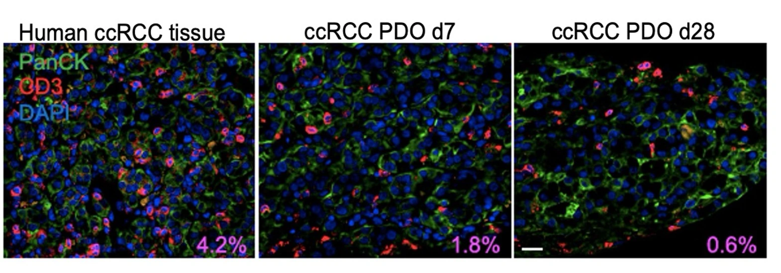
Model context: source tissue and PDO culture time points are shown together to compare tumor-epithelial and CD3-positive immune-cell signals before treatment design.
TRiCBIO ccRCC PDO 2.0 immune-context study data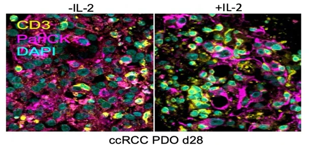 View image
Treatment context: imaging with and without IL-2 shows tissue and cellular changes under an immune-stimulation condition within the same study.
TRiCBIO ccRCC PDO 2.0 immune-context study data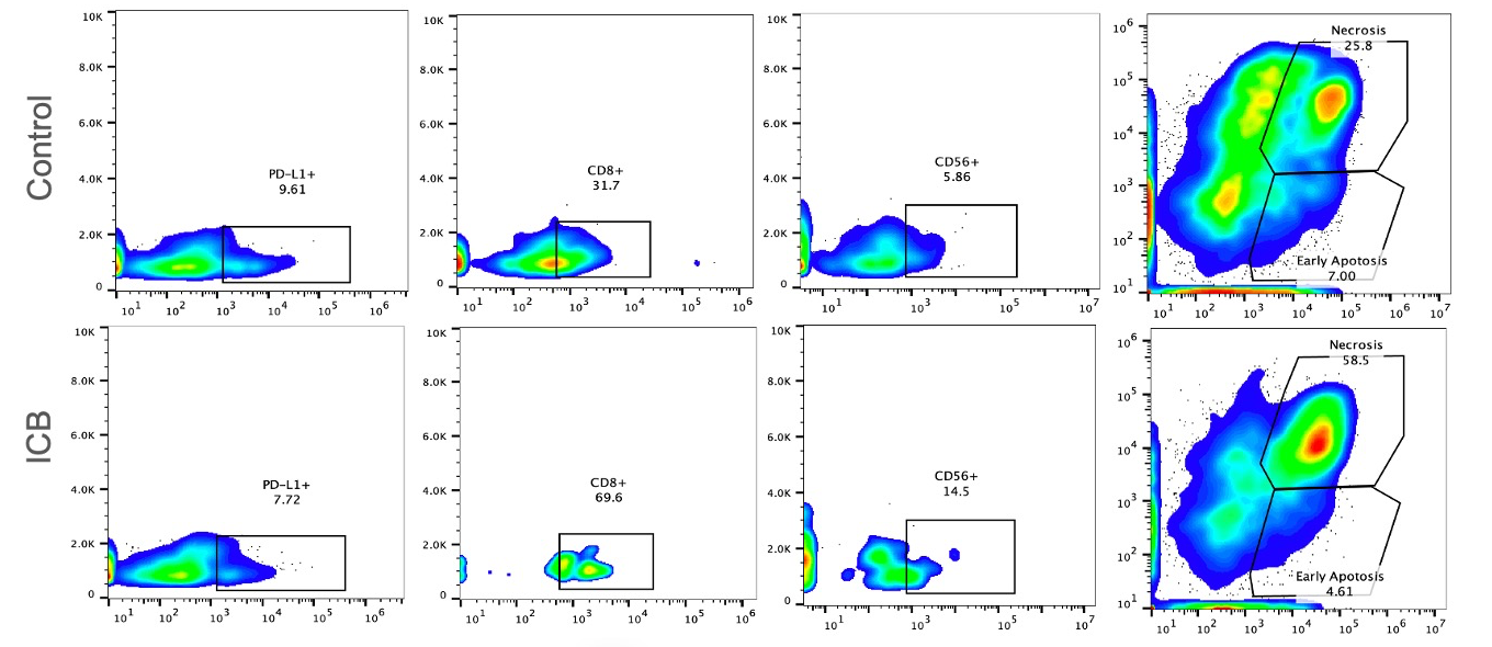 View image
Functional readouts: immune-associated cell phenotypes are interpreted alongside early-apoptosis and necrosis measurements after treatment in a multi-endpoint study.
TRiCBIO ccRCC PDO 2.0 immune-context study dataStudy process
From the study question to data and reporting
Sample & study goal
Confirm tissue source, mechanism and priority question
Model & QC
Document architecture, culture state and measurable components
Treatment
Set dose, timing, controls and combinations
Integrated assays
Interpret histology, flow, cytokine and functional readouts together
Results & report
Summarize key findings, applicable conditions and recommended follow-up studies
Further reading
Explore the science and plan your study
ALI foundation for tumor-immune studies
See how the Cell 2018 study informs ALI models, immune baselines and functional endpoints.
See program relevance and key figures ↗PDO 2.0 TME studies
Explore tissue imaging, T-cell profiling and treatment readouts from renal and lung cancer studies.
View study data ↗Models, assays and study delivery
See how model selection, experiments, interpretation and delivery fit into a project.
See the project workflow ↗Related models and services
Find relevant models and services
DOCUMENT REQUEST
Request the white paper and explore PDO 2.0 / ALI
Share a work email or phone number, with your research area if useful. The team will send the 2026 English edition (EN018) after review and can include relevant model or study materials.