
Establishes culture, tissue and marker baselines for candidate comparison or mechanism studies in a specific HCC model.
Model & assay dataCase study
HCC PDO and PDO 2.0: culture format and tissue identity
HCC PDO and PDO 2.0 studies combine culture morphology, source-tissue comparison, CD31 and the figure-labelled HEP-1 staining to guide efficacy and mechanism endpoints.
01Study objective02Model & design03Assays & findings04Decision & next step
Model & sampleHepatocellular carcinoma PDO and PDO 2.0 with source-tissue comparison
ObjectiveEstablish architecture, model identity and downstream study direction through complementary HCC model evidence
Study setupModel establishment, tissue characterization and treatment conditions selected for the study
AssaysBright-field, whole-mount immunofluorescence, H&E, CD31 and the figure-labelled HEP-1 staining
Study evidenceTRiCBIO model and assay data with study figures
DeliverablesDeliverables are defined by the study scope and may include images, assay results, an analysis summary, methods and a study report
CULTURE FORMAT & TISSUE IDENTITY
PDO morphology and HCC histology
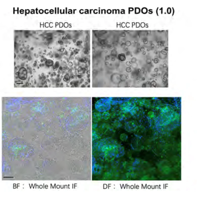
Bright-field and whole-mount immunofluorescence show culture morphology and cell state; HCC source-tissue and PDO H&E, CD31 and the figure-labelled HEP-1 staining then guide efficacy and mechanism endpoints.
 View image
Conventional PDO culture: bright-field and whole-mount immunofluorescence show model morphology and cell state, supporting characterization and downstream endpoint design.
Study data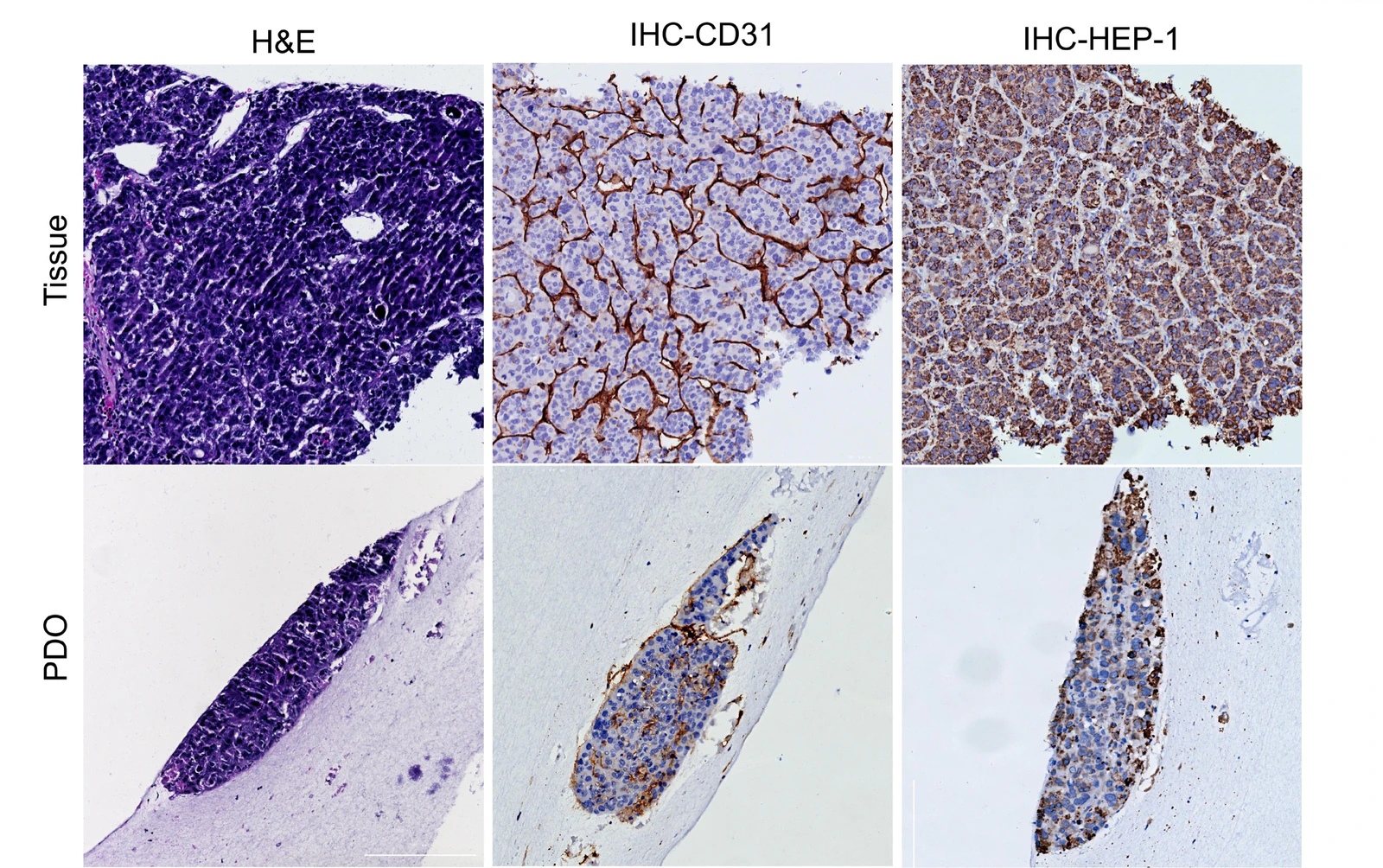 View image
HCC tissue and model profiling: paired H&E, CD31 and the figure-labelled HEP-1 immunohistochemistry in HCC source tissue and PDO show tissue architecture plus vascular and marker-staining context for selecting subsequent target and mechanism readouts.
Model & assay dataEstablish HCC model context from source-tissue comparison
HCC source-tissue morphology, PDO histology and overall model condition guide culture-format and endpoint selection across conventional PDO and PDO 2.0 studies.
Document PDO culture morphology and cell state
Bright-field and whole-mount immunofluorescence document 3D growth and cell state, then combine with source-tissue/PDO histology and the staining context visible in the figures to define the model profile used in treatment studies.
Compare histology and vascular staining context
Source-tissue and PDO H&E, CD31 and the figure-labelled HEP-1 immunohistochemistry establish tissue structure and staining context before efficacy or mechanism studies.
- Source-tissue comparison
- Figure-labelled HEP-1 staining
- Endothelial profiling
- Culture and histology QC
INTERPRETATION
Move from model identity into candidate and mechanism studies
Culture morphology, histology and the staining context visible in the figures are assessed with HCC pathology and can be extended through independent samples and functional endpoints.
Build treatment studies around target expression, dose, time and mechanism endpoints, then compare across independent samples.
Related models and services
Choose the right models and services for the next study
PROJECT DISCUSSION
Discuss a study built around your program
Tell us which model, assay or finding you want to extend. Our scientists will adapt the approach to your candidate, samples and decision point.